


The hip joint (HJ) is a complex joint formed by several bones: the femur, pubis, ilium, and ischium.Surrounded by a periarticular bursa and a strong muscle-ligament corset, protected by subcutaneous fat and skin.
The ilium, ischium and pubis form the pelvic bones and are connected by hyaline cartilage in the acetabulum.These bones fuse before the age of 16.
A characteristic feature of the femoral joint is the acetabulum structure which is only partially covered by cartilage, at the top and sides.The middle and lower segments are occupied by adipose tissue and femoral ligaments, covered by synovial membrane.
Reason
Pain in the hip joint can cause damage to the intra-articular elements or nearby structures:
- skin and subcutaneous tissue;
- muscles and ligaments;
- synovial bursa;
- acetabular lip (cartilaginous edge along the edge of the acetabulum);
- articular surface of the femur or pelvis.
Pain in the joint area is caused by inflammation or violation of the integrity of its constituent structures.Most often, pain occurs when infection enters the joint cavity (infectious arthritis) and autoimmune damage (rheumatoid and reactive arthritis).
Mechanical injuries are also common, resulting in damage to bone epiphyses, ligaments, synovial membranes, and other tissues.Active people and athletes who experience high levels of physical activity are more susceptible to injury.
Also at risk are elderly people who experience pain in the pelvic bones due to degenerative-dystrophic changes in the cartilage, as well as children and adolescents during times of hormonal changes.
Pain in the left or right hip joint is caused by metabolic diseases such as diabetes mellitus, pseudogout and obesity.
A complete list of possible diseases is as follows:
- Perthes disease;
- arthritis;
- Koenig's disease;
- diabetic arthropathy;
- pseudo gout;
- intermittent hydrarthrosis (intermittent dropsy of the joints);
- chondromatosis;
- reactive, rheumatoid and infectious arthritis;
- juvenile epiphysiolysis;
- injury.
Perthes disease
With Perthes disease, the blood supply to the femoral head is disrupted, leading to aseptic necrosis (death) of cartilage tissue.Most children under the age of 14, mostly boys, are affected.
The main symptom of Perthes disease is persistent pain in the hip joint, which gets worse when walking.Children often complain that their legs hurt starting from the hips and start to feel weak.
In the early stages, symptoms are mild, leading to a delay in diagnosis, when an impression (intra-articular) fracture has already occurred.The destructive process is accompanied by increased pain, swelling of soft tissues and stiffness of limb movements.The patient cannot rotate the hips outward, rotate, flex or straighten them.Moving the leg to the side is also difficult.
Disorders of the autonomic nervous system are also observed: the feet become cold and pale, and sweat profusely.Sometimes the body temperature rises to subfebrile levels.
Note: in Perthes disease, lesions can be unilateral or bilateral.In most cases, one of the joints suffers less and recovers more quickly.
arthritis
Osteoarthritis of the hip joint is called coxarthrosis and is diagnosed mainly in elderly people.This disease develops slowly, but causes irreversible changes.The pathological process begins with damage to the cartilage which becomes thinner due to an increase in the thickness and viscosity of the synovial fluid.
The development of coxarthrosis leads to joint deformation, muscle atrophy and significant limitation of movement up to complete immobility.The pain syndrome in arthrosis is undulating (not constant) and is localized on the outer side of the thigh, but can spread to the groin, buttocks and lower back.
In the second stage of arthrosis, painful sensations cover the inside of the thigh and sometimes reach the knee.As the disease progresses, the pain in the hip gets worse and sometimes subsides with rest.
Coxarthrosis can be primary and secondary.Primary coxarthrosis develops against the background of osteochondrosis or arthrosis of the knee.Prerequisites for secondary coxarthrosis may be hip dysplasia, congenital hip dislocation, Perthes disease, arthritis, and traumatic injuries (dislocations and fractures).
Koenig's disease
If the thigh on the side of the joint hurts, the cause may be death of cartilage tissue (necrosis) - Koenig's disease.This disease is most often found in young men aged 16-30 years, who complain of pain, decreased range of motion, and periodic “stuckness” in the legs.
Koenig's disease develops in several stages: first, the cartilage tissue softens, then hardens and begins to separate from the articular surfaces of the bones.In the third or fourth stage, the necrotic area is rejected and enters the articular cavity.This causes a buildup of effusion (fluid), stiffness of movement and blockage of the left or right joints.
Reference: the presence of a "joint mouse" in the hip joint causes the development of coxarthrosis.
Diabetic arthropathy
Osteoarthropathy, or Charcot joint, is observed in diabetes mellitus and is characterized by progressive deformation accompanied by pain of varying intensity.Pain sensations are expressed rather weakly or are completely absent, since in this disease the sensitivity is sharply reduced due to pathological changes in the nerve fibers.
Diabetic arthropathy occurs in long-term diabetes and is one of its complications.This occurs most often in women who do not receive complete or ineffective treatment.It should be noted that the hip joint is very rarely affected.
Pseudogout
As a result of impaired calcium metabolism, calcium crystals begin to accumulate in the joint tissue, and chondrocalcinosis, or pseudogout, develops.The disease received this name due to the similarity of symptoms with gout, which is characterized by its paroxysmal course.
Acute, sharp pain appears suddenly: the affected area becomes red and swollen, and becomes hot to the touch.The attack of inflammation lasts from several hours to several weeks, then everything goes away.With chondrocalcinosis, pain may occur in the left or right side of the pelvis.
In the majority of cases, pseudogout occurs without an obvious cause, and even examinations cannot detect calcium metabolism disorders.It is suspected that the cause of this disease lies in local metabolic abnormalities in the joints.In one in a hundred patients, chondrocalcinosis develops against the background of existing systemic diseases - diabetes, renal failure, hemochromatosis, hypothyroidism, etc.
Synovial chondromatosis
Chondromatosis of the joints, or cartilage island metaplasia of the synovium, primarily affects large joints, including the hips.Most often, this pathology occurs in middle-aged and elderly men, however, there are cases of congenital chondromatosis.
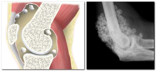
With chondromatosis, the synovial membrane degenerates into cartilage or bone tissue, resulting in the formation of chondromics or bone bodies up to 5 cm in size in the joint cavity.
The clinical picture of insular metaplasia is similar to arthritis: patients are bothered by pain in the hip bones, leg mobility is limited, and a characteristic crunching sound is heard when moving.
Since chondromatosis is a dysplastic process with the formation of chondromic bodies, the occurrence of “articular mice” cannot be excluded.In this case, the “mouse” may get stuck between the articular surfaces of the bones, which will cause partial or complete blockage of the joint.The joint remains blocked until the chondromic bodies enter the lumen of the capsule, and only after that movement is fully restored.
Help: frequent or prolonged joint congestion can trigger the development of coxarthrosis.Complications of synovial chondromatosis are stiffness (contractures) and muscle atrophy.
arthritis
Arthritis is localized inflammation of the articular surfaces of the acetabulum and femur.Damage to the hip joint is called coxitis, which is accompanied by a dull pain in the back of the thigh and groin.
There are several types of arthritis, the most common type affecting the hip joint is the infectious form.Other types are diagnosed less frequently.Why does infectious arthritis occur?The development of pathology begins after bacteria and viruses enter the joint cavity.
The clinical picture of infectious arthritis may differ depending on the type of causative microorganism.However, there are 5 typical signs that are observed in all patients:
- pain in the joints of the right or left leg (bilateral damage is also possible);
- swelling and swelling of the joints;
- redness of the skin;
- decreased motor abilities;
- increase in body temperature.
At the beginning of the disease, sufferers experience severe pain, especially when standing from a sitting position.His joints ached almost constantly;the pain makes it impossible to stand or sit.It should be noted that the infectious form of arthritis is always accompanied by fever, chills, headache, weakness and nausea.
Juvenile epiphysiolysis
The term epiphysiolysis literally means putrefaction, destruction of the articular surfaces of bones, or more precisely, the cartilage that covers them.A characteristic feature of this damage is the cessation of growth in bone length, which causes asymmetry in the lower extremities.
In adults, epiphysiolysis occurs when a fracture occurs with displacement or rupture of the epiphysis.Destruction of the epiphyses in the growth zone is possible only in adolescence, which is why the disease is called juvenile.
Juvenile epiphysiolysis is an endocrine-orthopedic pathology, which is based on an imbalance between growth hormone and sex hormones.These two groups of hormones are important for the normal function of cartilage tissue.
The dominance of growth hormone over sex hormone causes a decrease in the mechanical strength of the femoral growth zone, and displacement of the epiphysis occurs.The tip of the bone is located below and behind the acetabulum.
Typical symptoms of epiphysiolysis include pain in the right or left side of the thigh (depending on which joint is affected), lameness, and an unnatural position of the leg.The sore leg turns outward, the muscles of the buttocks, thighs and legs atrophy.
Treatment
To treat Perthes disease, chondroprotectors are prescribed to promote cartilage regeneration and angioprotectors necessary to improve blood circulation.Complex therapy also includes massage, exercise therapy, physiotherapy - UHF, electrophoresis with calcium and phosphorus, mud and ozokerite applications.
Patients with Perthes disease are advised to unload the limb and use an orthopedic device (cast), as well as a special bed to prevent deformation of the femoral head.
What to do and what medications to take for arthrosis depends on the stage of the disease.The following remedies help relieve pain and slow down the pathological process at stages 1-2:
- nonsteroidal anti-inflammatory drugs (NSAIDs);
- vasodilator;
- muscle relaxants to relax muscles;
- chondroprotector;
- hormonal (for severe pain);
- ointments and compresses with anti-inflammatory or chondroprotective effect.
At stage 3-4, patients are advised to undergo surgery.
Koenig's disease can only be treated surgically;during arthroscopic surgery, the affected area of cartilage is removed.
Treatment of diabetic arthropathy includes correction of the underlying disease – diabetes mellitus, wearing special bandages and taking medications.All patients, regardless of the stage of the disease, are prescribed antiresorptive drugs - bisphosphonates, as well as products with vitamin D and calcium.To eliminate pain and inflammation, drugs from the NSAID and corticosteroid groups are prescribed.If there are infectious complications, antibiotic therapy is performed.
There is no specific treatment for pseudogout;during exacerbations, anti-inflammatory drugs are prescribed.A large amount of fluid collected in the joint is an indication for intra-articular puncture, during which the fluid is pumped out and corticosteroid drugs are administered.
Chondromatosis of the hip joint requires mandatory surgical intervention, the volume of which depends on the scale of the lesion.If the number of chondromic bodies is small, they are removed by partial synovectomy (excision of the synovial membrane) or minimally invasive arthroscopy (through three punctures).Surgical treatment of progressive forms of chondromatosis can only be radical and carried out using open arthrotomy or complete (total) synovectomy.
Therapy for acute infectious arthritis includes the mandatory application of plaster to the hip joint area, taking medications of various groups (NSAIDs, antibiotics, steroids).When a purulent process develops, a series of medical punctures are performed to clean the joint.
Treatment of juvenile epiphysiolysis is only surgical.During surgery, closed bone repositioning is performed using skeletal traction.Then the combined bone sections are fixed with pins and grafts.
Absolutely all pathologies of the hip joint are serious diseases that require mandatory medical supervision.Any injury after a fall or impact that is accompanied by severe pain, limited mobility, and changes in joint configuration requires emergency medical attention.If there is no traumatic injury, but pain of varying intensity often occurs in the joint, you need to make an appointment with a therapist or rheumatologist and undergo an examination.